Objective
Detail the progress of an adolescent soccer player with right-sided chronic medial foot pain due to striking an opponent’s leg while kicking the ball. The patient underwent diagnostic ultrasound and a conservative treatment plan.
Clinical Features
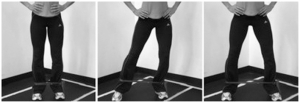
The most important features were hindfoot varus, forefoot abduction, flatfoot deformity, and inability to single leg heel raise due to pain. Conventional treatment was aimed at decreasing hypertonicity and improving function of the posterior tibialis muscle and tendon.
Intervention and Outcome
Conservative treatment approach utilized soft tissue therapy in the form of Active Release Technique®, and eccentric exercises designed to focus on the posterior tibial muscle and lower limb stability. Outcome measures included subjective pain ratings, and resisted muscle testing.
Conclusion
A patient with posterior tibialis tendonopathy due to injury while playing soccer was relieved of his pain after 4 treatments over 4 weeks of soft tissue therapy and rehabilitative exercises focusing on the lower limb, specifically the posterior tibialis muscle.
Introduction
Injuries are an unfortunate part of any sporting activity and soccer is no exception. Due to the nature of the sport the majority of soccer injuries occur in the lower limb, especially the ankle.1,2 It should come as no surprise that lower limb injuries are a common complaint that present to a chiropractic sports clinic. Injuries in soccer typically occur because of tackling, being tackled, running, shooting, twisting and turning, jumping and landing.2 A suggested reason for the higher incidences of ankle injuries is its close approximation to the ball, which is the main object of control in the sport. The chances of sustaining an ankle injury are highest when a player is tackling, shooting, or dribbling.2 Risk factors for soccer related injuries include age, gender, skill level, environment, and surface type.1 Epidemiological evidence indicates that approximately 10–15 injuries occur in soccer per 1000 playing hours.1 Older age players appear to be more prone to injury than younger ones, as do higher skill level athletes.2 Environment can also play a role in increased incidence, as playing indoors or on artificial turf has been linked with an increase in injury. Males are more susceptible to ankle injuries while it is reported that knees are the most frequent injury for females.2
Avulsion injuries for soccer players are rare. They can present as groin pain from a sports hernia (tear of the abdominal muscles attachments to the pubic tubercle), or rectus femoris injury (tear of proximal attachment from the anterior inferior iliac spine avulsion).3,4 In younger athletes consideration must also be given to apophyseal injuries. These injuries typically occur between 8 and 15 years of age and are associated with skeletal immaturity, muscle-tendon imbalance, and repetitive microtrauma. Common apophyseal injuries in young athletes include Osgood-Schlatter disease (tibial tuberosity), Sever’s disease (posterior calcaneus), Sindig-Larsen-Johansson syndrome (inferior patella), and medial epicondylitis (medial humeral epicondyle).5 Navicular fractures can also occur in athletes though they are far less common in soccer players.6,7 As well to-date there have been few avulsion injuries reported with the navicular bone (main distal attachment site of the posterior tibialis muscle) and not a single navicular apophsyeal injury in a young athlete.8,9
A common tendon injury associated with soccer however is the posterior tibial tendon (PTT).1 The prevalence of PPT dysfunction in the general population is approximately 3.3% and often results in adult acquired flatfoot deformity.10 In soccer athletes injuries to this tendon result from repetitive kicking of the ball. During a ball strike the ankle is forced into excessive plantar-flexion and the foot into extreme pronation. Consequently the PTT can become irritated due to friction against the medial malleolus and subject to a number of pathological changes such as avulsion, longitudinal tear, or rupture of the mid tendon substance.1,11 The medial malleolus acts to change the direction of pull by the tendon. This is believed to cause increased stress on the tendon as ruptures typically occur in this location.12
The mechanism of injury is theorized to be a degenerative process of the PTT caused by decreased blood supply. This then results in a tendon dysfunction with possible rupture and gradual collapse of the medial longitudinal arch of the foot. Other common findings associated with PTT dysfunction include hindfoot valgus deformity and forefoot abduction. Mechanically the PTT is stressed during gait immediately after heel strike as the hindfoot moves from eversion into inversion. As the angulation of the hindfoot increases the forefoot compensates by progressively moving into an abducted position. Cutting sports such as soccer are known to place increased stress on the PTT.13,14 Additionally, as the PTT becomes dysfunctional there is an imbalance with its antagonist muscle the fibularis brevis. The dominating unopposed action of the fibularis brevis also contributes to the deformities mentioned above (medial longitudinal arch collapse, hindfoot valgus, and forefoot abduction).15,16 Finally, the presence of a prominent navicular tuberosity or an accessory navicular have been linked to PTT tears. Again the proposed mechanism of injury is chronic irritation of the tendon. However, the location of the tear is now at the navicular insertion, not at the level of the medial malleolus.

This really answered my problem, thank you!